
At Cell Systems, we strive to create the most biological, most physiologically-relevant conditions for your research. Our antibody-free primary cells are ideal tools for optimizing your experiments. Not only is it hard to find a more biologically-relevant cell type, but our deep inventory guarantees access to the same cell population for the entirety of your research program. This means more consistency and increased reproducibility.
To create even more relevant experimental systems, we recently partnered with 4Dcell, a biotech company working to revolutionize cell culture by controlling the cellular microenvironment.
4Dcell has developed cell culture products to confine cells to limited spaces and shapes, better mimicking the in vivo microenvironment. A more realistic cell microenvironment means more predictive modeling in early phase R&D, and a deeper understanding of cellular mechanism and function in the human body.
Why is cellular confinement important? In vivo, cells are highly organized to form structures, tissues, and organs – and that structure helps determine function. For example, smooth muscle cell alignment around the small intestine yields a tissue capable of exerting substantial force. Conventional 2D cell culture conditions – in which cells are given a boundless surface on which to grow in any orientation – don’t reflect the cell’s natural microenvironment. Our recent experiments provide evidence that high quality primary cells can organize well within these confined spaces.
4Dcell’s micropatterns, microchannels, and cell confinement tools offer a simple way to begin your investigations of cellular function within the native microenvironment. Contact our R&D team to discuss applications for cell confinement in your research.
Visit www.cell-systems.com for information and ordering of our antibody free primary cells, media and reagents.
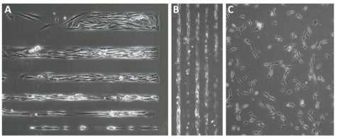
ACBRI 716 Primary Human Aortic Smooth Muscle Cells cultured on 4Dcell micropatterned slides (Panels A and B) versus standard tissue culture dish (Panel C). Each surface was coated with AttachmentFactorTM extracellular matrix and all cultures were maintained in Complete Classic Medium with Serum and CultureBoostTM. Phase contrast images captured 2hr after plating. Magnification: Panel A, 20X; Panels B and C, 10X.