

3D models are often applauded for being more predictive than 2D cultures, but not all scientists know how to maximize their 3D models’ biological relevance to reach their full potential.
Using primary human cells (derived directly from human tissues) can greatly enhance the biological relevance of your models and steer your experiments in the right direction from the start.
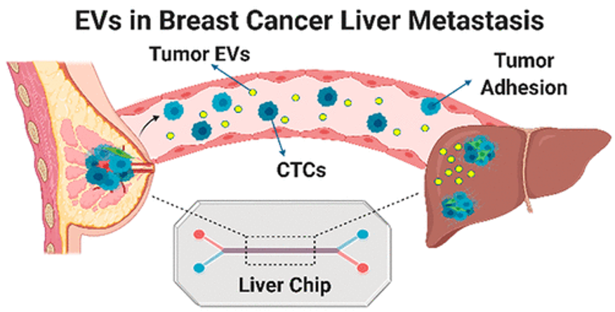
In a study by Kim et al., researchers use primary liver sinusoidal endothelial cells (Cat# ACBRI 566) to construct a 3D microfluidic human liver-chip to better understand how breast cancer-derived extracellular vehicles (EVs) can influence metastasis in the liver. While there have been many studies done on breast cancer, the relationship between its EVs and metastasis is still not well understood.
The metastatic tumor microenvironment constructed in previous research lack some of the features that this study has.
For instance, the use of unadulterated primary human cells preserves the true characteristics of cells in their in vivo state. This allows scientists to eliminate non-relevant variables such as the genetical and phenotypical differences observed in immortalized cell lines. Additionally, 3D microfluidic device allows scientists to model the flowing bloodstream that is sometimes neglected in past studies.
With the model’s biological relevance, not only can this liver-chip of primary cells allow scientists to better understand breast-cancer metastasis, but it can be used to emulate other cancer-specific metastasis in the future as well.
If you are interested in maximizing your models’ biological relevance with primary liver sinusoidal endothelial cells (Cat# 566) or other unadulterated primary human cells, please visit www.cell-systems.com or contact customerservice@cell-systems.com.