These antibody-free human primary cells were isolated by enzymatic dissociation of normal human cerebral cortex tissue without the use of positive selection of antibodies.
Primary Human Brain Pericytes Cells (ACBRI 498)

- PRODUCT INFO
- CITATIONS
- TESTS
- RESEARCH
- DOCUMENTATION
Increase Biological Relevance with Human Primary Cells
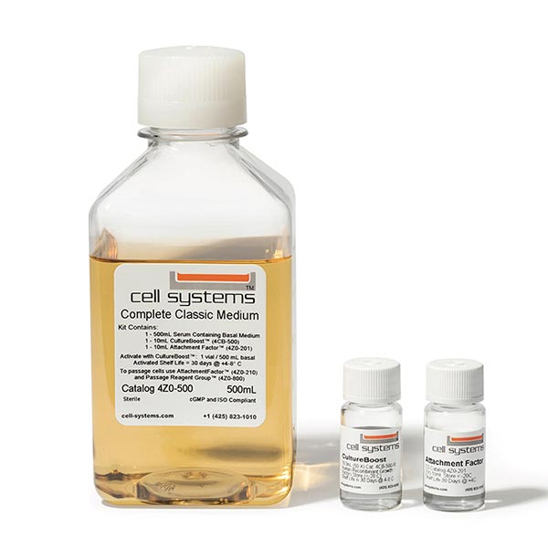
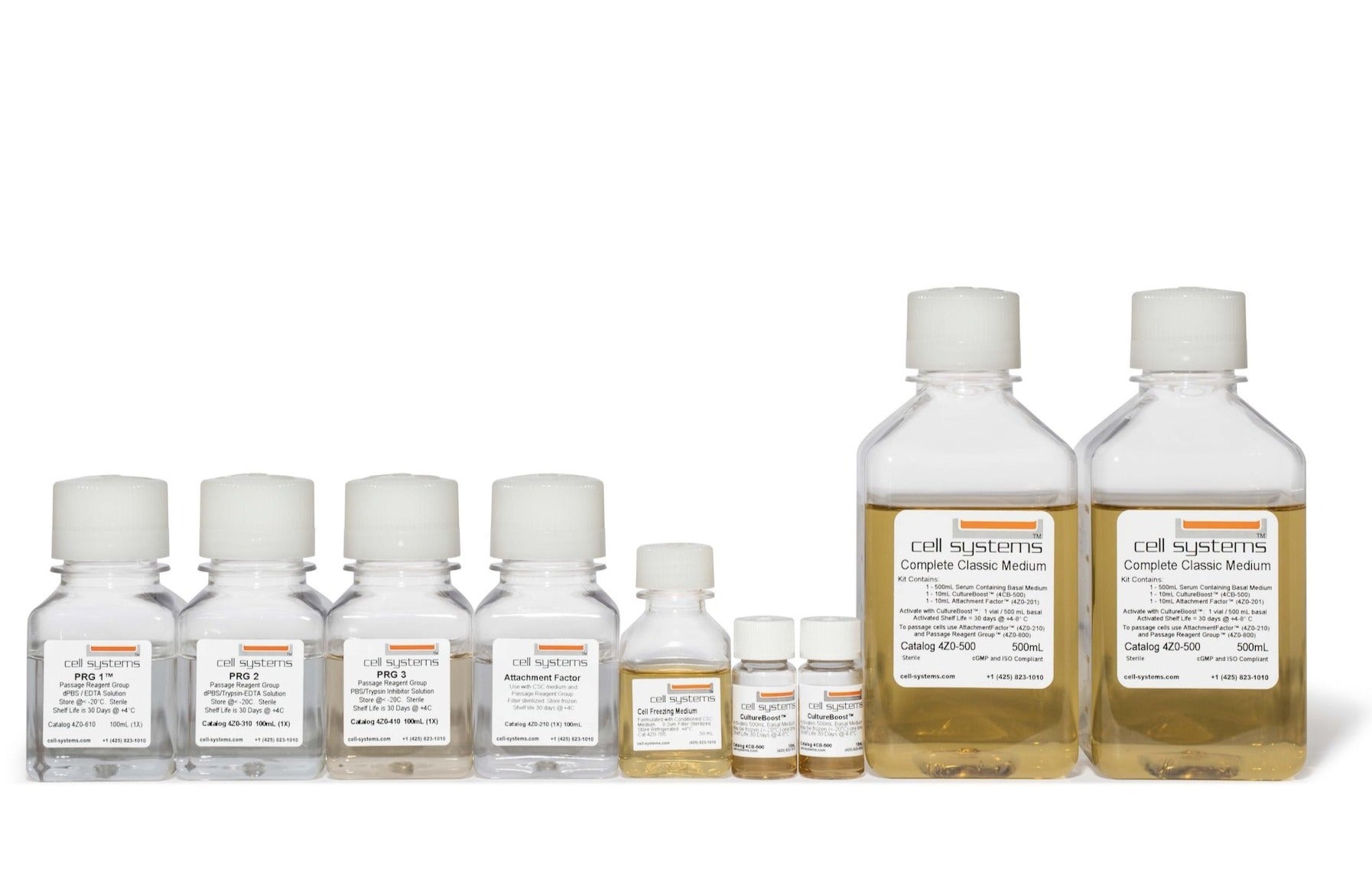
Our antibody-free primary cells can offer a more biologically relevant cell culture tool for scientists to enhance their research insights. These primary cells were originated using Cell Systems Complete Serum-Free Medium (SF-4Z0-500) and subsequently grown and passaged in Cell Systems Complete Classic Medium (4Z0-500). The cells are cryopreserved in Cell Systems Cell Freezing Medium (4Z0-705).
- Isolated from normal, healthy donor tissue
- Available at Passage 3 (<12 cumulative population doublings)
- Each vial contains approximately 1 x 10⁶ cells
- Supplied as frozen, cryopreserved 1 mL vials or as actively proliferating flask
- Available in reserved lots to enhance consistency in your research program
Each vial of cells is shipped to customers with 1mL of Bac-Off® and .25mL of CultureBoost™.
Cell Profile for ACBRI 498: Surface Markers, Genes & Soluble Molecules (Show more>)
| Cell Marks, Genes & Molecules Tested | Methodologies | Most Cited Studies |
| RAGE variant proteins |
| |
| Pericytes’ effects on endothelial production of G-CSF, IL-6, IL-8 and IL-17 in chips and transwell cultures |
| |
| PDGFR-β, β-Actin, Akt, NGF, BDNF, NT-3 |
| |
| Fibronectin +, Vimentin, CD68, NG-2
|
|
For details on our primary human cell profiles, contact us at customerservice@cell-systems.com
ELEVATE RESEARCH CONSISTENCY
WITH OUR RESERVED LOT PROGRAM
ELEVATE RESEARCH CONSISTENCY
WITH RESERVED LOT PROGRAM
The Reserved Lot Program is designed to assist researchers in securing consistent supplies of human primary cells for their long-term research projects.
How the LOT Program Can Benefit You:
- Reserve a Specific Number of Vials: You can reserve a set number of vials from a designated lot at no cost, ensuring that you have continued access to the same consistent cell lot throughout your research.
- Request New Lots: Our LOT program allows you to request the generation of new lots, with up to 200 vials made specifically for your research needs. While we typically offer 1.00 x 10⁶ cells/vial, we can customize the cell count per vial if necessary to meet your requirements.
RESERVE YOUR LOT TODAY! Email us at customerservice@cell-systems.com or call 425-823-1010.
A Selection of Citations for ACBRI 498 from Scientific Journals
Discover additional research on Google Scholar that utilizes Cell Systems primary cells.
- "A quasi-physiological microfluidic blood-brain barrier model for brain permeability studies". Noorani and Bhalerao et al. Pharmaceutics, 2021.
- "PDGFR-β restores blood-brain barrier functions in a mouse model of focal cerebral ischemia". Shen and Xu et al. J Cerebral Blood Flow & Metabolism, 2018.
- "Distinct contributions of astrocytes and pericytes to neuroinflammation Identified in a 3D human blood-brain barrier on a chip". Herland et al. PLoS One, 2016.
- "Retinal pericytes and cytomegalovirus infectivity: implications for HCMV-induced retinopathy and congenital ocular disease". Wilkerson et al. J Neuroinflammation, 2015.
- "Brain vascular pericytes following ischemia have multipotential stem cell activity to differentiate into neural and vascular lineage cells". Nakagomi et al. Stem Cells, 2015.
- "Inflammation-induced endothelial cell-derived extracellular vesicles modulate the cellular status of pericytes". Yamamoto et al. Scientific Reports, 2015.
- "Infection and upregulation of proinflammatory cytokines in human brain vascular pericytes by human cytomegalovirus". Alcendor et al. J Neuroinflammation, 2012.
- "PDE4 regulates tissue plasminogen activator expression of human brain microvascular endothelial cells". Yang et al. Thrombosis Research, 2012.
- "Expressions and roles of AMIGO gene family in vascular endothelial cells". Hossain et al. Intl J Bioscience Biochemistry Bioinformatics, 2012.
- "PDGF receptor β signaling in pericytes following ischemic brain injury". Arimura et al. Current Neurovascular Research, 2012.
- "Neurotrophin production in brain pericytes during hypoxia: A role of pericytes for neuroprotection". Ishitsuka et al. Microvascular Research, 2012.
Standard Tests
| TESTS | RESULTS |
| HIV Serologic Test (donor level HIV AB EIA) | Negative |
| RPR Syphilis Test | Negative |
| Hepatitis B (HBV) and Hepatitis C (HCV) PCR Test (at frozen cell level) | Negative |
Retail Production (P3)Tests
| TESTS | RESULTS |
| Bacterial Sterility (culture method) by independent lab | Pass |
| Fungal Sterility (culture method) by independent lab | Pass |
| Mycoplasma Sterility (culture method) by independent lab | Pass |
Cell Markers and Functional Tests
| MARKER | RESULT |
| Desmin pericyte marker | > 98% positive by immunofluorescence at P3 |
| PDGFR-b pericyte marker | > 98% positive by immunofluorescence at P3 and P10 |
| NG2 pericyte marker | > 98% positive by immunofluorescence at P3 and P10 |
| CD13 pericyte marker | > 98% positive by immunofluorescence at P3 and P10 |
| a-SMA pericyte marker | > 98% positive by immunofluorescence at P10 |
| CD31 endothelial cell marker | < 2% by immunofluorescence at P3 and P10 |
| Uptake of Di-I-Ac-LDL, endothelial cell test | < 2% by immunofluorescence at P3 and P10 |
| MAP2 neuronal marker | < 2% by immunofluorescence at P3 and P10 |
| Neurofilament neuronal marker | < 2% by immunofluorescence at P3 and P10 |
| S100A4 fibroblast marker | < 2% by immunofluorescence at P3 and P10 |
| GFAP astrocyte marker | < 2% by immunofluorescence at P3 and P10 |
| GS astrocyte marker | < 2% by immunofluorescence at P3 and P10 |
| CD11b microglial marker | < 2% by immunofluorescence at P3 and P10 |
| IbaI microglial marker | < 2% by immunofluorescence at P3 and P10 |
Immunofluorescence Imagery and Characterization in Peer-Reviewed Literature
ACBRI 498 at Passage 3 labeled with antibodies against pericyte markers desmin, PDGFR-b, NG2, and CD13.
From Herland and van der Meer et al. (2016) PLoS ONE 11(3): e0150360.
Fig 2. Co-culture of human brain microvascular endothelial cells (ACBRI 376), pericytes (ACBRI 498) and astrocytes in the 3D BBB chip.
Schematic illustrations of the cells populating the 3D vessel structures for the three experimental set-ups are shown at the top, and fluorescence confocal micrographs of the engineered brain microvessel viewed from the top (A, D, G) or shown in cross-section at either low (B, E, H) or high (C, F, I) magnification (rectangles in lower magnifications images indicate respective areas shown at higher magnification below). The fluorescence micrographs show the cell distributions in 3D BBB chips containing brain microvascular endothelium alone (A-C), endothelium with prior plating of brain pericytes (ACBRI 498) on the surface of the gel in the central lumen (D-F) or endothelium with brain astrocytes embedded in the surrounding gel (G-I). High-magnification cross-sections are projections of confocal stacks (bars, 200 μm in A,B,D,E,G,H and 30 μm in C, F, I). Green indicates F-actin staining, blue represents Hoechst-stained nuclei, and magenta corresponds to VE-Cadherin staining, except for G where morphology and intensity masks were used to discriminate astrocytes (green) from endothelial cells (ACBRI 376) (magenta); original image can be seen in S2 Movie. Arrows indicate contact points between endothelium and pericytes (ACBRI 498) (F) or astrocytes (I).
From Herland and van der Meer et al. (2016) PLoS ONE 11(3): e0150360.
Fig 4. Establishment of a low permeability barrier by the engineered brain microvascular endothelium in the 3D BBB chip.
A) Fluorescence micrograph of a chip containing a cylindrical collagen gel viewed from above with (left) or without (right) a lining endothelial (ACBRI 376) monolayer after five days of culture (left; Cell-lined lumen) compared to a chip with an empty collagen lumen (right). The images were recorded at 0 (top) and 500 (bottom) sec after injection of fluorescently-labeled 3 kDa dextran to analyze the dynamics of dextran diffusion and visualize endothelial barrier function in the 3D BBB chip. Note that the presence of the endothelium significantly restricts dye diffusion compared to gels without cells (left versus right). B) Apparent permeabilities of the endothelium cultured in the 3D BBB chip calculated from the diffusion of 3 kDa dextran with an endothelial (ACBRI 376) monolayer (Endo; n = 6), an endothelial (ACBRI 376) monolayer surrounded by astrocytes (Endo+Astro; n = 3) and an endothelial (ACBRI 376) monolayer surrounded by pericytes (ACBRI 498) (Endo+Peri; n = 3). Error bars indicate S.E. M.; * p<0.05, Student’s t-test.
COMPANION PRODUCTS:
OPTIMIZE YOUR RESEARCH
WITH HUMAN PRIMARY CELLS
Avoid the obstacles from immortalized cell lines or animal models and gain more pertinent insights into human cell biology.
Cell Systems cells are available for in vitro research purposes only and may not be transferred out of the direct control of the recipient Institution/Agency/Organization. Cell Systems cells may not be genetically altered in any way without prior written permission from Cell Systems. Use of Cell Systems materials (evidenced by placement of any order for product) constitutes knowledge, understanding and binding acceptance of these restrictions on behalf of the recipient Institution/Agency/Organization.
Cell Systems was created to further the knowledge of eukaryotic cell biology through laboratory research, publications and teaching. Cell Systems provides cells and cell culture products to other research entities - public and private.


